Table of Contents
Preface
Introduction
The Sonny Burke Story
Chapter I What is Thoracic Outlet Syndrome? (TOS)
Chapter II Anatomy
Chapter III The TOS Controversy
Chapter IV History, Cause, and Patient Presentations
Chapter V Physical Examination Findings
Chapter VI Diagnostic Tests
Chapter VII Standard of Care Approaches – Surgical and Non-Surgical
Chapter VIII Frequently Asked Questions
Chapter IX Case Histories of Patients
Chapter X The Human Spring Approach to Treatment and Prevention

Chapter II
![]()
Anatomy of Thoracic Outlet Syndrome
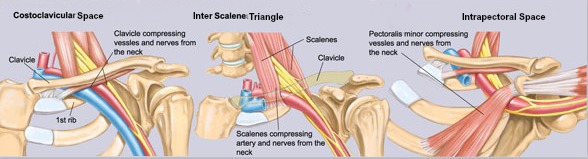
Thoracic Outlet Syndrome – Three Potential Areas of Compression
Thoracic outlet syndrome is the often misdiagnosed cause of neck pain, shoulder pain and arm disability. It is thought to be neurovascular compression seen at the thoracic outlet, which is something that anatomists can’t agree on. The actual name doesn’t properly describe the condition.
Thoracic Outlet Areas Of Compression
3 areas of potential regions of compression consisting of:
- The Inter Scalene Triangle
- The Costoclavicular Space
- The Intrapectoral Space
The Inter Scalene Triangle
This is bordered by the anterior and middle scalenes. The supraclavicular bundle consisting of the subclavian vein, the subclavian artery and the brachial plexus emanate from this triangle and it’s an area where any one of these structures can become compressed and cause symptomatology.
The Costoclavicular Space
The area below the clavicle and above the first rib represents the costoclavicular space. Few patients and doctors understand that the ribs actually go up this high at the face of the neck.
The Interpectoral Space
The last space is the interpectoral space and that is in the area of pectoralis minor and that area can be an area of compression.
Arteries, Veins and Nerves pass through the Thoracic Outlet
Doctors have to be aware of these multiple areas of compression and have an understanding of what symptoms can be related to each one of these areas so they can better treat the patient. The three-neurovascular structures that pass through the thoracic outlet area are the brachial plexus consisting of cervical nerves C5, C6, C7, C8 and T1. The subclavian artery is the artery that supplies the arm with blood, oxygen and nutrients. The subclavian vein drains the blood away from the arm and back to the heart.
What is Venous Thoracic Outlet Syndrome?
Venous thoracic outlet syndrome (TOS) is also known as Paget-Schroetter syndrome or subclavian vein effort thrombosis. Paget determined that the symptoms of the upper extremity (ie, arm swelling) were a result of subclavian vein thrombosis. Von Schroetter further proposed that the upper extremity venous symptoms were a result of thrombosis of the subclavian vein at the thoracic outlet. At the level of the thoracic outlet, the subclavian vein passes over the first rib, anterior to the insertion of the anterior scalene muscle. This space is called the costoclavicular space and is located between the clavicle and subclavius muscle, superior to the subclavian vein with the first rib being inferior to the subclavian vein.
Venous TOS is a result of extrinsic compression of the subclavian vein, which results in injury of the vein, and eventual, stenosis (narrowing) and thrombosis (clotting). The most common causes of extrinsic compression of the subclavian vein are a narrow costoclavicular space or muscular hypertrophy of the subclavius or anterior scalene.

A symptom of Venous Thoracic Outlet Syndrome is pitting edema of the arm
The symptoms of venous TOS are caused by subclavian vein thrombosis and/or stenosis. The symptoms involve the upper extremity (arm), and include: arm swelling, arm heaviness or aching of the arm, and cyanosis. An individual may notice prominent, distended veins in the upper chest and shoulder region as well as pitting edema of the arm (pictured above), distending veins and dilated collateral veins. Especially after activities which require repetitive use of the involved extremity. Rarely, a pulmonary embolism may occur.
Paget–von Schrötter disease, is a form of deep vein thromboisis (DVT), in which blood clots form in the veins of the arms. These DVTs typically occur in the axillary or subclavian veins.

READ MORE POSTS ON THORACIC OUTLET SYNDROM

www.thoracicoutletsyndrome.com
WATCH VIDEOS OF PRESENTATIONS BY DR STOXEN ABOUT TOS ON THE TEAM DOCTORS THORACIC OUTLET SYNDROME YOUTUBE CHANNEL